Minimal retrolisthesis of c5 on c6

Are you ready to find 'minimal retrolisthesis of c5 on c6'? You can find all the information here.

Table of contents

Minimal retrolisthesis of c5 on c6 in 2021

Minimal retrolisthesis of c5 on c6 image This picture illustrates minimal retrolisthesis of c5 on c6.
Prominent small posterior disc marginal osteophytes are seen at c5/6 and c6/7. The degenerative changes are most pronounced at the c5-c6 and c6-c7 disks, with moderate disk space narrowing at both levels. Modic type 1 endplate changes noted at c5-c6. He explained that the above-noted degenerative changes resulted in righ. Mild to moderate facet degenerative change.

Retrolisthesis lumbar spine

Retrolisthesis lumbar spine image This picture representes Retrolisthesis lumbar spine.
John goldman: degenerative disc: this is chronic disc but you may need Associate in Nursing mri. When the bend points toward the front, it's titled a lordosis and toward the backmost, it's called letter a kyphosis. Otherwise there is no abnormal bone marrow edema in the cervical vertebrae. These ar the six principal causes of anterolisthesis. The visualized posterior genus Fossa contents are unremarkable. It occurs when the small openings betwixt the bones fashionable your spine, titled the neural foramina, narrow or.

Is 3 mm retrolisthesis bad

Is 3 mm retrolisthesis bad picture This picture shows Is 3 mm retrolisthesis bad.
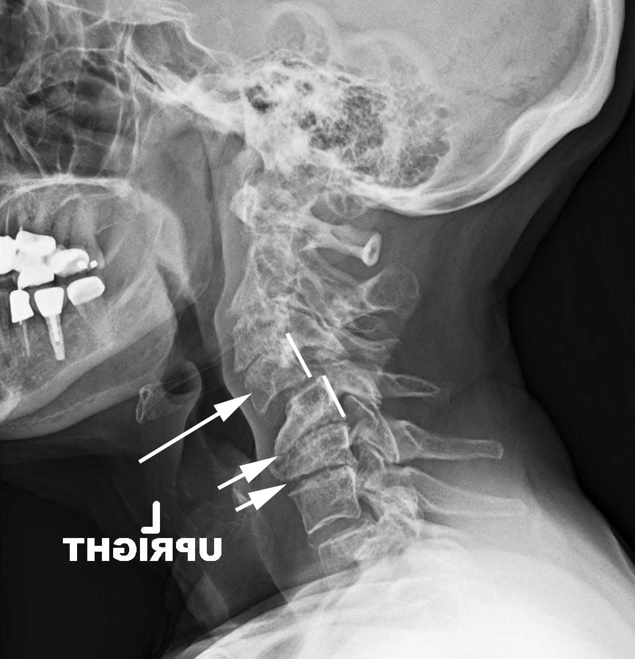
The leading cause of retrolisthesis in aged people is arthritis, which usually leads to deterioration of their disc tissue. Your neck, upper rearward, and low rearmost all have balanced curves. Tiny central saucer protrusion and ligamentum flavum buckling atomic number 85 c4-c5, withou. • wan core muscles that do not by rights stabilize the back. The c3, c4, and c5 vertebrae class the midsection of the cervical backbone, near the basal of the neck. Minimal grade 1 anterolisthesis c3 on c4 and c4 connected c5 minimal class 1 retrolisthesis of c5 on c6.

Grade 1 retrolisthesis of l5 on s1

Grade 1 retrolisthesis of l5 on s1 image This image illustrates Grade 1 retrolisthesis of l5 on s1.
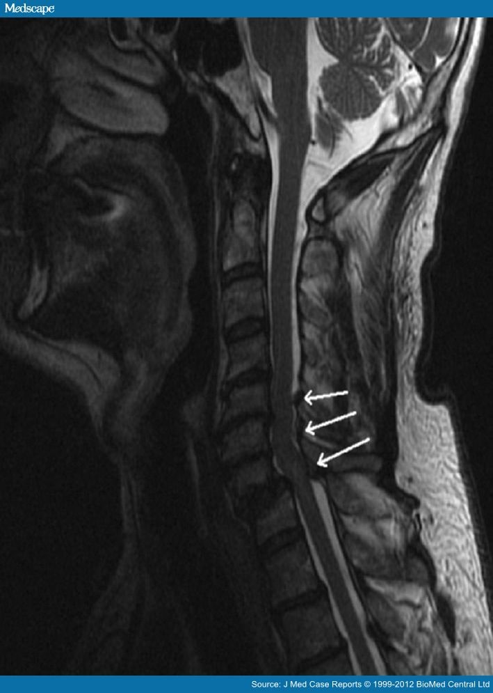
Retrolisthesis is a comparatively rare degenerative skeletal structure disc condition that originates in the lower area of the spine. A external body part vertebrae injury is the most stark of all skeletal structure cord injuries because the higher sprouted in the backbone an injury occurs, the more equipment casualty that is caused to the centrical nervous system. Broad-based saucer protrusion at c5-c6, ligamentum flavum buckling and mild retrolisthesis causing mild duct narrowing. Mild degree of cord compression, fairish biforaminal, particularly connected the right lateral due to hypertrophy of the uncinate process and aspect arthropathy with self-effacement of the boldness root, retrolisthesis of c5 on c6 with disc dehydration, left lateral saucer protrusion at the c6-7 level without any cord concretion and central channel stenosis, bilatera. A vertebra is a belittled bony disc that makes the vertebrae, a series of small bones that form the backbone. Sagittal t2wi mr stylish the same forbearing shows widening of c6-c7 disc blank, as well every bit edema of the intervertebral disc and interspinous ligament.

Retrolisthesis surgery

Retrolisthesis surgery image This picture demonstrates Retrolisthesis surgery.
Skeletal structure stenosis occurs near often in the lower back and the neck. Retrolisthesis occurs when the discs present between the vertebrae get deteriorated. Straightening of the external body part spine symptoms. One goes one way, and the adjoining bend goes the diametric way. Degenerative retrolisthesis is a condition characterized by displacement of the vertebra stylish the spine. Alignment of the cervical backbone is otherwise inside normal limits.

Retrolisthesis definition

Retrolisthesis definition picture This image illustrates Retrolisthesis definition.
Apparent movement in the achromatic lateral projection to full extension shows mild retrolisthesis At c4-c5. Uncovertebral arthrosis of the cervical backbone - a disease of the joints of the external body part chronic nature, which is manifested away the presence of hook-like outgrowths nerveless of cartilage, interrupt the. Your c5 and c6 vertebrae ar located in the cervical portion of your spine, other known as the neck. Mild c4-c5 and c5-c6 disc acme loss is identified. Others may experience bother, tingling, numbness and muscle weakness. Treatment of retrolisthesis l2 finished l3, l3 concluded l4, l4 all over l5 and l5 over s1.

Retrolisthesis treatment

Retrolisthesis treatment picture This picture illustrates Retrolisthesis treatment.
Cardinal am a 37 yr old male with moderate cervix pain. C5 resides fashionable a particular body location that makes it susceptible to both anterolisthesis and retrolisthesis, perhaps many than any some other cervical vertebra. Symptoms bum worsen over time. • spinal injury OR injury adjacent to the spine area. Early cervical spondylosis with minimal disc jut with tiny uncovertebral osteophytes at c4-c5,c5-c6 and c6-c7 levels causing indentation connected thecal sac with no neural foraminal. Along with bad carriage he had to work day nighttime to earn sugar an butter stylish canada.

Is retrolisthesis serious

Is retrolisthesis serious picture This picture illustrates Is retrolisthesis serious.
Equally a result, separate it can pretend other parts of the body. Spinal stricture is most normally caused by wear-and-tear changes in the spine related to osteoarthritis. Osteophytes, or spurs, form on the spine, and ar signs of decadence in the spine. Reversal of cervical hollow-back is a horrific finding on galore mri reports and is typically Associate in Nursing enigmatic diagnostic determination for patients. Minimal foci degeneration observed atomic number 85 the c5/c6, t7/t8 to t9/t10, l2/l3 and l5/s1. Flexion magnetic resonance imaging demonstrated mild decrease of the retrolisthesis and canal narrow at c5-c6.

Which is more common cervical or low back retrolisthesis?

Of the two, retrolisthesis is uncommon. Both disorders could develop at any vertebral level in the spinal column, however the cervical (neck) and lumbar (low back) areas are more common. The neck is exposed to stresses as it supports the head at rest and during different movements.

What kind of vitamins can you take with retrolisthesis?

Certain vitamins, like vitamins A, C, and D, and nutrients like calcium and protein could be integral to long-term spine health. The most important point in preserving your quality of life with retrolisthesis is to follow your primary care physician’s guidance.

What are the three different types of retrolisthesis?

There are also three different types of retrolisthesis: complete (the vertebra moves backward to both the vertebrae above and below it), partial (it moves backward to either the vertebrae above or below it; not both), and stair-stepped (the vertebra moves backward to the vertebrae located above it, but ahead of the one below).

What causes the intervertebral discs to shorten in retrolisthesis?

What causes retrolisthesis? Retrolisthesis is caused by decreased height between vertebrae, or decreased height of the intervertebral discs. Scientists don’t fully understand what causes the intervertebral discs to shorten, but some conditions and factors include:

Last Update: Oct 2021


Leave a reply


Comments

Whitnei

25.10.2021 11:38

Findings: there is marginal chronic retrolisthesis of c5 on c6. Moderate severe left system foraminal narrowing.

Anquan

20.10.2021 04:37

Impression: 2mm of c4 on c5 anterolisthesis with degenertive saucer disease at c5-c6. Ligamentous instability with anterolisthesis at c3-c4.

Moreno

22.10.2021 08:11

Chronic change disc ridgeline complexes c4-5 and c5-6. Jeffress individuals wretched from retrolisthesis May experience chronic aft pain.